
Sebum Analysis Facial Imaging: How It Works, What It Measures & How Accurate It Is
Seeing shiny skin is easy. Measuring facial sebum is much harder. Surface oil can vary by facial region, cleansing history, temperature, humidity, cosmetics, hydration, and time of day, so a visual inspection cannot reliably tell you how much sebum is present or how it is distributed. Sebum analysis facial imaging uses controlled optical imaging and image analysis to document surface-lipid patterns across the face, offering spatial information that point-based measurements may not provide.
However, an important distinction comes first: an image signal is not automatically the same thing as an absolute chemical measurement of sebum. The usefulness and accuracy of facial imaging depend on the optical method, calibration, image processing, reference standard, and measurement protocol. Research has demonstrated that fluorescence imaging can map facial sebum distribution and that image-derived fluorescence can correlate with conventional sebum measurements, while also highlighting the importance of standardized acquisition and interpretation. :contentReference[oaicite:0]{index=0}
This guide explains how facial sebum imaging works, what it actually measures, how it compares with a Sebumeter, which variables can distort results, and how professionals can choose an imaging system based on the measurement objective rather than marketing claims.
What Is Sebum Analysis Facial Imaging?
Sebum analysis facial imaging is the use of specialized photography, optical illumination, spectral imaging, or image-processing techniques to visualize and analyze skin-surface lipid patterns. Depending on the system, the resulting image may be assessed for signal intensity, fluorescent regions, coverage, distribution, or other image-derived characteristics associated with surface sebum.
The key advantage is spatial information. Instead of obtaining a measurement from one small contact area, imaging can document multiple facial regions simultaneously and preserve a visual record for comparison. Research on digital fluorescent facial imaging has specifically explored automated analysis of sebum distribution as an alternative to some limitations associated with point measurements and subjective visual assessment. :contentReference[oaicite:1]{index=1}
Sebum vs. Sebaceous Gland Activity
These terms should not be treated as interchangeable. Sebum is the lipid-rich material present on the skin surface, while sebaceous gland activity refers to secretion by the glands. Surface accumulation can be influenced by secretion, spreading, removal, skin temperature, sweat, and other factors. Traditional research distinguishes a static casual sebum level from dynamic measures such as sebum excretion rate. :contentReference[oaicite:2]{index=2}
What Facial Imaging Can and Cannot Tell You
Facial imaging can reveal patterns in optical signals associated with surface conditions. It can help identify where oil-related fluorescence or contrast is concentrated and can provide standardized documentation for comparison.
It does not automatically prove how much lipid is present in absolute mass units, nor does it directly establish the secretion rate of an individual sebaceous gland. A strong image signal should therefore be interpreted as an image-derived measurement or proxy unless the specific imaging system has been validated against an appropriate reference method.
Imaging-Based Analysis vs. Visual Skin Assessment
Unaided observation is affected by ambient light, viewing angle, skin tone, cosmetics, hydration, and observer judgment. Standardized imaging can reduce some of these sources of variation by fixing acquisition conditions and preserving the same facial regions for subsequent comparison.
That makes imaging particularly useful when the goal is objective documentation, spatial mapping, consultation support, or longitudinal monitoring rather than simply deciding whether skin looks oily.
How Does Sebum Analysis Facial Imaging Work?
A professional facial imaging workflow generally moves from controlled image acquisition to optical signal detection, region segmentation, and interpretation. The exact process varies substantially between devices, so the following describes the measurement logic rather than claiming that every AI skin analyzer uses the same sebum-specific technique.
1. Standardize the Face and Capture Conditions
The subject is positioned consistently while illumination, camera distance, exposure, facial angle, and other acquisition settings are controlled. This matters because changes in lighting or camera settings can change pixel intensity even when the underlying skin has not changed.
For longitudinal monitoring, consistency is especially important. A baseline captured under one set of conditions should ideally be compared with follow-up images acquired using the same protocol.
2. Capture Optical Signals Associated With Surface Sebum
Different imaging systems exploit different optical interactions. Fluorescence imaging can use ultraviolet or other excitation light to reveal fluorescent signals associated with skin-surface components. Published research has used digital fluorescent imaging to analyze facial sebum distribution, while later research has investigated automated segmentation of UV-induced facial fluorescence. :contentReference[oaicite:3]{index=3}
Polarized-light techniques have a different purpose. Cross-polarized imaging can suppress certain surface reflections and improve visualization of selected skin features, but it should not automatically be described as a direct sebum assay.
Multispectral imaging extends the concept by capturing information across multiple wavelength bands. A larger spectral dataset can help separate or characterize optical differences that ordinary RGB photography cannot resolve, but the resulting indicators still require appropriate validation before they are treated as quantitative biological measurements.
3. Segment Relevant Facial Regions
Image analysis can divide the face into standardized regions of interest, such as the forehead, nose, cheeks, and chin. Automated segmentation can then identify pixels or areas meeting specified signal criteria.
This is important because facial sebum is not uniformly distributed. The T-zone often differs from the cheeks, and research using fluorescence imaging has demonstrated region-specific facial patterns associated with sebum levels. :contentReference[oaicite:4]{index=4}
4. Convert Image Signals Into Quantitative or Semi-Quantitative Results
Software may calculate signal intensity, affected area, percentage coverage, regional differences, or comparative scores. These values can make an image easier to track over time.
But a critical measurement principle applies: image intensity is not automatically equivalent to absolute sebum mass. A quantitative-looking number is only as meaningful as the calibration and validation behind the algorithm. Even conventional Sebumeter output requires careful interpretation and calibration when researchers want to relate readings to actual quantities of oil. :contentReference[oaicite:5]{index=5}
What Does Facial Sebum Imaging Actually Measure?
Surface Sebum Quantity and Relative Oiliness
Some imaging approaches can estimate differences in surface lipid accumulation. In practical professional use, this can help distinguish relatively oilier and less oily facial zones.
The safest interpretation is usually comparative: one region has more detected signal than another, or a standardized follow-up image shows a change from baseline. Absolute mass claims require validation against an appropriate reference method.
Sebum Distribution Across the Face
Spatial distribution is one of facial imaging's strongest advantages. Instead of reporting a single forehead or cheek measurement, an imaging system can create a broader map showing where oil-associated signals are concentrated.
This can be valuable during professional skin analysis because clients may have combination patterns rather than uniformly oily or dry skin.
Sebum-Associated Fluorescence or Optical Contrast
Fluorescent signals can provide useful information about sebum-related facial patterns. Studies have reported relationships between UV-induced red fluorescence, casual sebum levels, and particular facial distributions. :contentReference[oaicite:6]{index=6}
Nevertheless, fluorescence is an optical signal. Its intensity can depend on the imaging system, illumination, camera response, biological fluorophores, skin characteristics, and processing method. It should therefore be interpreted within the validated context of the specific protocol.
Sebaceous Gland Activity: What Imaging Does Not Directly Measure
A facial image showing more surface oil does not, by itself, establish that sebaceous glands are producing oil at a particular rate. Surface accumulation and glandular secretion are related but distinct endpoints.
Facial Sebum Imaging Methods Compared
Fluorescence Imaging
Fluorescence imaging captures emitted light after excitation. UV-induced fluorescence has been studied for facial sebum and follicular assessment, and image segmentation can convert fluorescent patterns into measurable regions. :contentReference[oaicite:7]{index=7}
Its major strength is visualization of spatial patterns that are difficult to capture through simple visual inspection. Its limitation is that fluorescence is influenced by the biological and optical properties of the skin and therefore requires controlled interpretation.
Polarized-Light and Cross-Polarized Imaging
Polarization changes how reflected light is captured. Cross-polarized imaging can reduce certain glare effects and make some pigmentation, redness, and texture differences easier to visualize.
It is important not to equate improved skin visualization with direct sebum quantification. A polarized image may support broader facial analysis without being a validated measurement of skin-surface lipid mass.
UV Imaging
UV-based imaging can reveal fluorescence patterns that are difficult to see under ordinary illumination. Research has examined UV-induced facial fluorescence in relation to sebum and acne-related features. :contentReference[oaicite:8]{index=8}
UV imaging requires appropriate equipment, controlled exposure, and safety considerations. The resulting fluorescence should also be interpreted carefully because different fluorescent components can contribute to the observed signal.
Multispectral Imaging
Multispectral imaging captures information from multiple wavelength bands rather than relying solely on conventional visible-light photography. This can create a richer optical dataset for assessing different facial characteristics.
However, more spectral channels do not automatically mean higher accuracy for every measurement. The relevant question is whether the wavelengths, algorithms, calibration, and reference measurements are validated for the endpoint being assessed.
Image-Based Sebum Scoring and Segmentation
Modern image analysis can identify pixels or facial regions according to predefined thresholds or machine-learning models. Research has demonstrated the feasibility of automated segmentation for UV-induced facial fluorescence, while also showing that algorithm performance can vary depending on signal strength and dataset characteristics. :contentReference[oaicite:9]{index=9}
For professional systems, software consistency can be just as important as camera resolution. A high-resolution image does not automatically produce a reliable skin measurement if region selection, thresholding, calibration, or interpretation is inconsistent.
Facial Imaging vs. Sebumeter: Which Is More Accurate?
There is no universal winner because the two approaches answer somewhat different questions.
How a Sebumeter Measures Skin Surface Lipids
A Sebumeter is a contact-based photometric instrument commonly used to assess skin-surface sebum. A measurement cassette or sampling surface interacts with the skin, and changes in optical properties are converted into a reading.
Research has shown that Sebumeter output can correlate with the amount of oily material deposited on skin, but the relationship is material-dependent and the output should not automatically be treated as a direct mass measurement without suitable calibration. :contentReference[oaicite:10]{index=10}
What Imaging Does Better
- Maps facial distribution instead of focusing on a single sampling point.
- Creates a visual record for consultation and documentation.
- Can compare multiple facial regions in one acquisition.
- Supports longitudinal image comparison when capture conditions are standardized.
- Can combine sebum-related information with broader facial characteristics when the system supports multispectral or AI analysis.
What a Sebumeter Does Better
- Provides a standardized localized measurement approach.
- Is well established in cosmetic and dermatological research.
- Can be useful when a defined point measurement is the primary endpoint.
- Can support quantitative studies when the instrument and protocol are appropriately calibrated.
Why “More Accurate” Is the Wrong Universal Question
Accuracy should be judged against the intended endpoint. If the objective is spatial mapping, facial imaging has an inherent advantage. If the objective is a localized quantitative measurement using an established contact method, a Sebumeter may be more appropriate.
For research or clinical claims, the strongest approach may involve using imaging for spatial information while validating or supplementing image-derived results with an established reference measurement.
Comparison Matrix: Imaging vs. Sebumeter
| Measurement consideration | Facial imaging | Sebumeter-style measurement |
| Spatial mapping | Strong | Limited to sampled locations |
| Visual documentation | Strong | Limited |
| Localized quantitative assessment | Depends on validation | Established use |
| Longitudinal image comparison | Strong when standardized | Possible through repeated point measurements |
| Susceptibility to protocol variation | Lighting, camera settings, cosmetics, positioning, and processing can matter | Sampling pressure, surface preparation, timing, and material response can matter |
| Best suited to | Mapping, visualization, consultation, and comparative monitoring | Standardized localized sebum measurement |
A Standardized Sebum Imaging Measurement Workflow
Reliable facial imaging depends less on taking a visually impressive photograph and more on repeating the same measurement conditions. Research on skin-surface lipid assessment emphasizes controlled temperature, humidity, acclimatization, and collection procedures. :contentReference[oaicite:11]{index=11}
Step 1: Control Skin Preparation
Record whether the subject has recently washed the face or applied moisturizer, sunscreen, makeup, oil, or another topical product. Residual products can alter optical signals and may also contribute their own oily materials to the skin surface.
For repeat assessments, use the same preparation protocol whenever possible.
Step 2: Control Temperature and Humidity
Allow the subject to acclimatize to the measurement environment and record relevant environmental conditions. Temperature can affect sebum spreading, while sweat and hydration can alter surface characteristics. :contentReference[oaicite:12]{index=12}
Step 3: Standardize Lighting and Camera Position
Use consistent facial positioning, camera distance, angle, exposure, and illumination. If a system uses specialized spectra, keep those acquisition parameters fixed for baseline and follow-up images.
Step 4: Define Facial Regions of Interest
Establish repeatable regions such as the forehead, nose, chin, and cheeks. Automated or guided segmentation can reduce inconsistency in selecting measurement areas.
Step 5: Capture Baseline and Repeat Measurements
Where the purpose is progress tracking, capture a baseline and repeat the same protocol at later visits. Multiple captures can also help identify whether an apparent difference is larger than ordinary acquisition variation.
Step 6: Record Protocol Conditions With the Result
Keep a record of skin preparation, time since cleansing, room conditions, device settings, and analysis method. Without this information, a numerical or visual difference between two sessions can be difficult to interpret.
What Factors Can Affect Facial Sebum Imaging Results?
Cleansing and Skin Preparation
Washing removes surface lipids and changes the starting condition of the skin. Measurements taken immediately after cleansing should not automatically be compared with measurements taken after several hours of normal activity.
Cosmetics, Sunscreen, and Skincare Residue
Topical products can create optical signals or alter the amount and distribution of material on the skin surface. Research evaluating Sebumeter measurements has also demonstrated that different oily materials can produce different instrument responses, reinforcing the need to distinguish sebum from topical product residue. :contentReference[oaicite:13]{index=13}
Hydration and Skin Barrier Condition
Hydration and sebum are different skin parameters. A person can have relatively high surface oil and still have altered stratum-corneum hydration. Combining complementary measurements can therefore provide a more useful profile than treating “oily” and “hydrated” as the same characteristic.
Corneometry is generally used to assess stratum-corneum hydration, while TEWL measures water movement through the skin and is commonly used as an indicator related to barrier function. These measurements should not be substituted for sebum measurement. :contentReference[oaicite:14]{index=14}
Ambient Lighting and Device Settings
Image-derived measurements can change when exposure, illumination, camera response, or positioning changes. This is particularly important when comparing signal intensity across different sessions.
Temperature, Humidity, Sweat, and Recent Activity
Heat, humidity, exercise, and sweating can change the surface environment. Earlier research on sebum measurement specifically identifies temperature and sweat as factors that can influence surface sebum behavior. :contentReference[oaicite:15]{index=15}
Time Since Cleansing and Time of Day
Skin-surface lipids change over time, and research has identified circadian variation as a consideration in sebum studies. For longitudinal work, keeping the timing of measurements reasonably consistent improves interpretability. :contentReference[oaicite:16]{index=16}
For broader professional skincare education and device-based care, the Skin and Beauty Care Devices collection provides a useful context for understanding how facial analysis tools fit into a wider skincare workflow.
How Accurate and Repeatable Is Sebum Analysis Facial Imaging?
The correct question is not simply whether an AI skin analyzer or imaging system is “accurate.” Instead, ask accurate for what endpoint, compared with which reference, under what protocol, and in which population?
Accuracy vs. Repeatability vs. Reproducibility
Accuracy concerns how closely a measurement approaches an accepted reference value. Repeatability concerns consistency under the same conditions. Reproducibility concerns consistency when relevant conditions, operators, sessions, or environments change.
A system may produce highly repeatable image scores without those scores being validated as absolute sebum mass. Conversely, a validated measurement method can still produce variable results if the protocol is poorly controlled.
Calibration and Reference Standards
Calibration connects an instrument's signal to a defined reference. For imaging, this can involve controlled optical references, standardized image acquisition, or comparison against an established measurement technique.
Research into Sebumeter quantification illustrates the principle: different oily materials produced different instrument responses, meaning calibration is necessary when quantitative claims depend on relating instrument output to actual material quantities. :contentReference[oaicite:17]{index=17}
Image Processing and Segmentation Variability
Algorithms may use thresholds, color characteristics, spectral signatures, region-of-interest definitions, or machine-learning segmentation. Changing these parameters can change the detected area.
Recent research on deep-learning segmentation of UV-induced facial fluorescence illustrates why model validation matters: performance differed between algorithms, particularly in weak or absent fluorescence regions, and the researchers emphasized the need for broader datasets and clinical-grade validation. :contentReference[oaicite:18]{index=18}
Device-Specific Validation
Do not transfer validation from one imaging platform to another simply because both use terms such as “AI,” “multispectral,” “UV,” or “8-spectrum.” The exact camera, illumination, software, population, acquisition protocol, and measurement endpoint matter.
How to Judge an Accuracy Claim
- What reference method was used?
- Was the imaging system calibrated?
- What endpoint was actually measured?
- How many subjects were included?
- Were skin tones and relevant populations adequately represented?
- Was repeatability reported?
- Were environmental conditions controlled?
- Were cosmetics and skincare residues controlled?
- Was the exact device and software version evaluated?
- Does the validation support absolute measurement, relative scoring, or visual screening?
Sebum Imaging vs. Other Skin Measurement Technologies
Sebum Imaging vs. Corneometry
Corneometry primarily assesses changes associated with water content in the stratum corneum. It is therefore a hydration measurement rather than a direct measurement of surface sebum.
Sebum Imaging vs. Transepidermal Water Loss (TEWL)
TEWL measures water loss through the skin. It is commonly used in research involving skin-barrier function and should not be interpreted as a measurement of facial oil. :contentReference[oaicite:19]{index=19}
Why Hydration and Sebum Should Be Evaluated Separately
Skin condition is multidimensional. Sebum describes a surface-lipid characteristic; hydration describes a different physiological property; TEWL provides information about water loss and barrier behavior. Treating all three as interchangeable can produce misleading skincare conclusions.
Combining Imaging With Multiple Biophysical Measurements
For research, formulation testing, or advanced professional assessment, combining spatial facial imaging with validated point measurements can provide more information than relying on one metric alone. The appropriate combination depends on the question being investigated.
Professionals building a broader device-based workflow can also explore the Body Care and Health Devices collection when considering how facial analysis fits alongside other non-invasive care technologies.
When Should Professionals Use Facial Sebum Imaging?
Professional Skin Consultations
Facial imaging can make consultations more structured by giving professionals a visual record of skin characteristics and regional differences. Instead of relying exclusively on subjective descriptions such as “your skin looks oily,” an imaging report can support a more systematic discussion of observed patterns.
Treatment Planning and Progress Tracking
Standardized images can document changes between consultations. This is particularly useful when the goal is comparative monitoring rather than diagnosing a medical condition.
Cosmetic Product and Formulation Research
Imaging can be useful in controlled product evaluations when researchers need spatial information about facial changes. Stronger claims still require a validated endpoint and appropriate study design.
Clinical or Research Applications
When imaging results are intended to support clinical research, regulatory claims, or scientific conclusions, the evidence requirements become more demanding. A professional aesthetic imaging device should not automatically be described as a clinical diagnostic instrument simply because it uses AI or multiple wavelengths.
When a Traditional Sebum Measurement Is Preferable
A direct, localized measurement can be preferable when the research question specifically requires a standardized point-based sebum value. Imaging is generally more compelling when spatial distribution, visual documentation, or multiple facial characteristics are important.
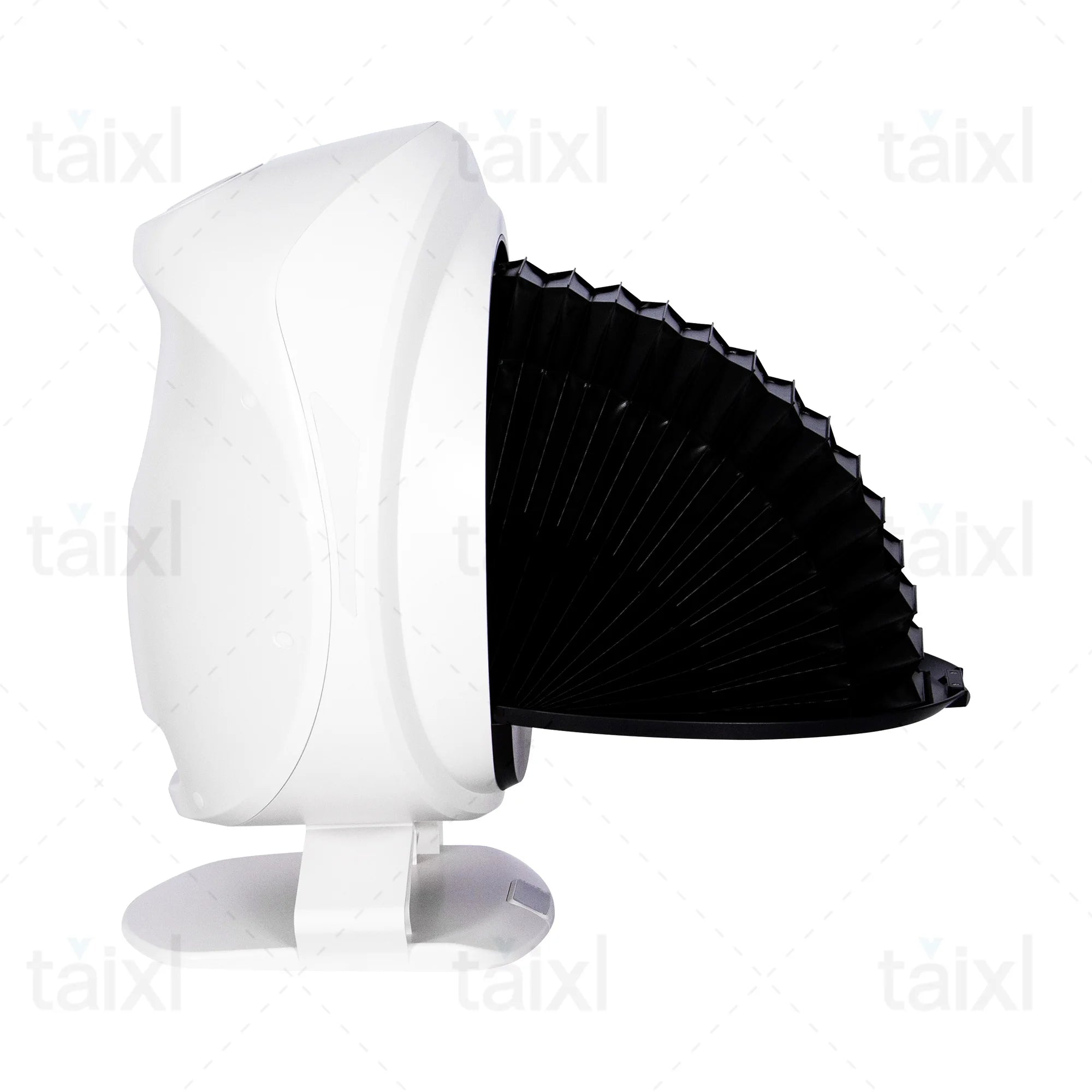
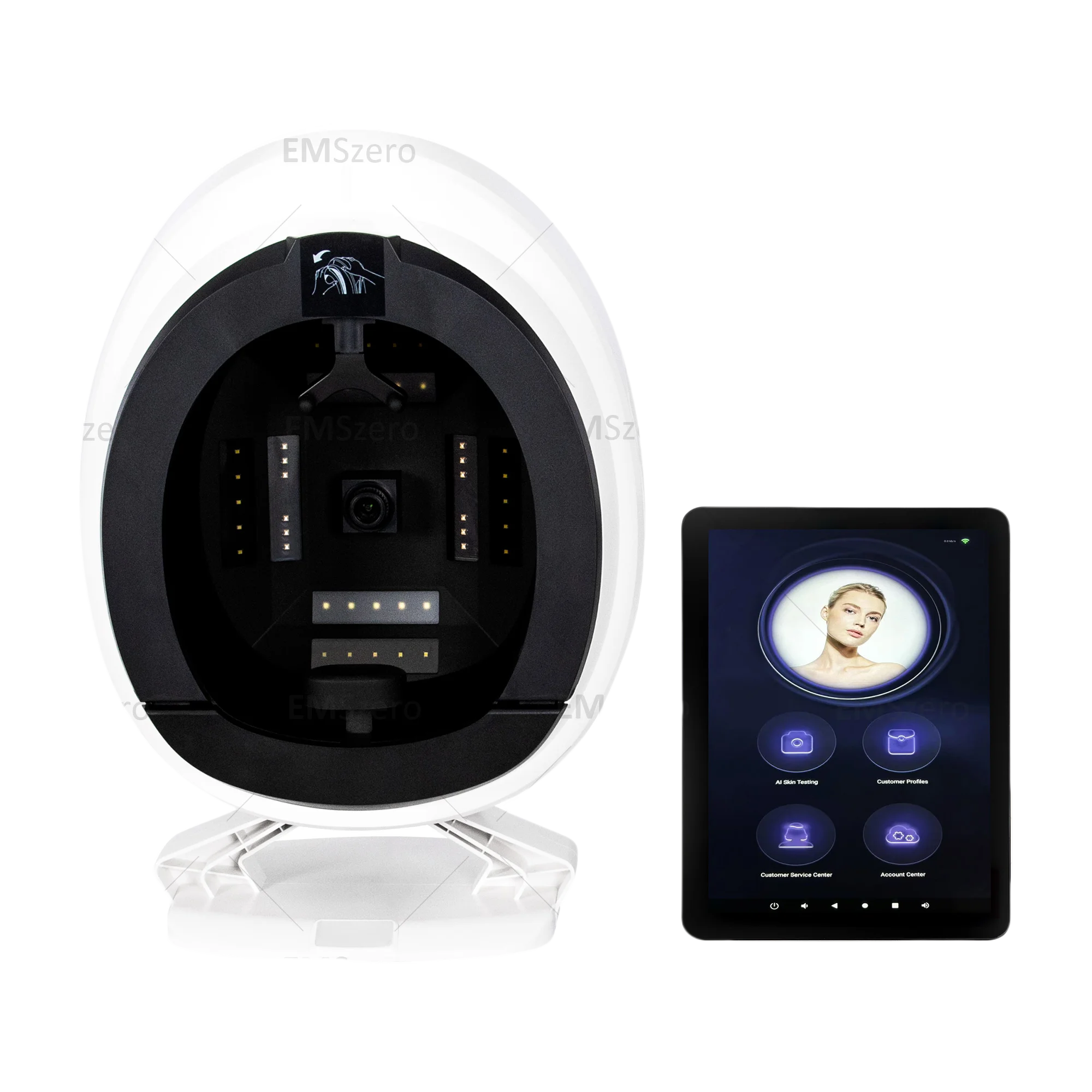
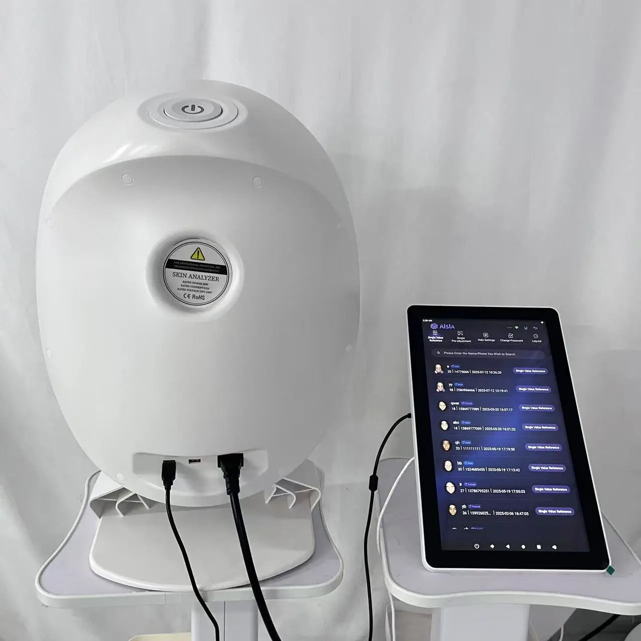
For professional salons and skincare businesses that want broader facial documentation rather than a standalone oil measurement, an AI Skin Analyzer for Professional 3D Facial Skin Analysis can serve as an example of a workflow-oriented system. The MYOSLIM device uses a 36MP industrial HD camera, 8-spectrum imaging, and AI-powered reporting, with the product specification indicating analysis of sebum alongside acne, pigmentation, pores, wrinkles, moisture, skin tone, and elasticity.
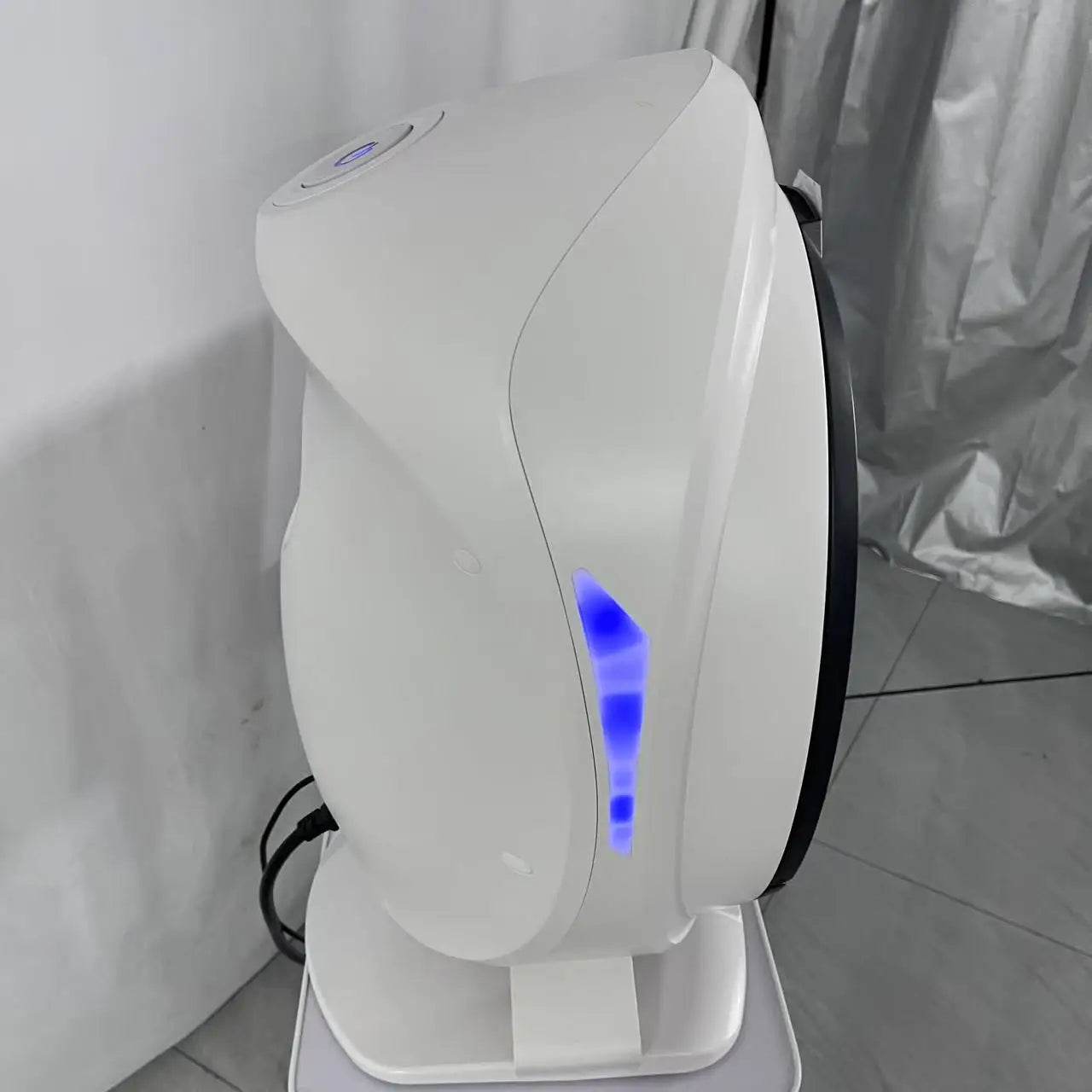
Its role should be understood in that context: a professional facial analysis system can help organize imaging and consultation data, but device-specific claims about absolute sebum quantification or clinical diagnostic accuracy should be based on validation evidence for the exact system and endpoint.
How to Choose a Facial Sebum Imaging System
Start With the Measurement Objective
First decide whether you need screening, spatial mapping, consultation visualization, longitudinal documentation, research measurement, or validated quantitative sebum assessment. Different objectives can justify different technologies.
Check the Imaging Modality and Optical Specifications
Look beyond megapixels. Consider the illumination wavelengths, spectral bands, polarization capabilities, camera consistency, exposure control, field of view, and whether the system can reproduce the same acquisition conditions from session to session.
Evaluate Calibration and Software
Ask how the software identifies facial regions, whether regions of interest can be standardized, how scoring is generated, whether algorithms can be configured, and whether reports or raw images can be retained for longitudinal comparison.
Look for Validation and Repeatability Evidence
Prefer evidence tied to the exact device rather than generic statements about AI skin analysis. A credible validation study should explain its reference method, population, protocol, endpoint, sample size, and measurement error.
Consider Workflow and Client-Facing Usability
Professional equipment also needs to work in the real world. Capture speed, positioning, hygiene, report generation, data management, image storage, and ease of explaining results to clients can matter as much as technical specifications.
Decision Matrix: Which Method Fits Which Goal?
| Primary goal | Preferred approach | Main priority | Typical application |
| Map facial oil distribution | Facial optical or fluorescence imaging | Spatial consistency | Professional consultation |
| Measure localized surface sebum | Validated Sebumeter-style method | Standardized point measurement | Research or controlled testing |
| Track multiple visible skin characteristics | Multispectral or AI facial imaging | Repeatable imaging and reporting | Salon or skincare clinic workflow |
| Assess hydration | Corneometry or another validated hydration method | Appropriate physiological endpoint | Skin research and product testing |
| Assess barrier-related water loss | TEWL instrumentation | Controlled environmental protocol | Clinical and cosmetic research |
| Support scientific quantitative claims | Validated reference method plus appropriate imaging | Calibration and validation | Research and claims substantiation |
For a salon or skincare clinic evaluating professional facial analysis equipment, the Body & Face Blog can provide additional context around facial-care technologies and professional workflows.
When a system needs to document more than sebum alone, the professional AI facial skin analyzer described above combines high-resolution imaging with 8-spectrum analysis and digital reporting. Its stated capabilities cover sebum as one indicator within a broader facial assessment rather than positioning the device as a standalone laboratory sebum assay.
Limitations and Common Misinterpretations
An Image Is Not Automatically a Direct Chemical Measurement
Optical imaging measures light behavior. If the objective is to determine the exact chemical composition or absolute mass of surface lipids, additional validated analytical methods may be necessary. Modern research into skin-surface lipids, for example, uses specialized sampling and analytical techniques to characterize lipid composition, demonstrating how different measurement questions require different methodologies. :contentReference[oaicite:20]{index=20}
More Visible Oil Does Not Always Mean Higher Sebum Production
Surface appearance can reflect accumulation and spreading, not simply secretion rate. Temperature, sweat, cleansing, topical products, and the physical properties of surface lipids can influence what is observed.
Do Not Diagnose Skin Conditions From Sebum Images Alone
A sebum-related image can contribute to professional screening and documentation, but an optical pattern should not be treated as a standalone medical diagnosis. Clinical diagnosis requires appropriate assessment by qualified healthcare professionals.
Standardization Matters More Than a Single Attractive Image
A visually impressive image is not necessarily a scientifically reliable measurement. For tracking changes, consistent preparation, positioning, illumination, software processing, and timing are more important than simply producing a high-resolution photograph.
Practical Checklist for Reliable Sebum Imaging
Before Imaging
- Record time since facial cleansing.
- Control or document skincare, sunscreen, makeup, and other topical products.
- Allow the subject to acclimatize to the measurement environment.
- Keep temperature and humidity reasonably consistent.
- Document unusual factors such as recent exercise or sweating.
During Imaging
- Use consistent facial positioning.
- Maintain a fixed camera distance and angle.
- Keep illumination and exposure settings consistent.
- Use the same facial regions of interest for repeat measurements.
- Capture repeat images when measurement reliability is important.
After Imaging
- Apply the same segmentation and analysis protocol.
- Distinguish image-derived scores from absolute sebum measurements.
- Record acquisition conditions with the result.
- Compare like-for-like baseline and follow-up images.
- Use validated reference measurements when quantitative claims require them.
- Avoid diagnosing skin disease from imaging results alone.
Frequently Asked Questions
How does sebum analysis facial imaging work?
It uses controlled optical imaging to capture facial signals associated with surface conditions, followed by image processing that can identify regions, calculate signal characteristics, and generate comparative or semi-quantitative results. Fluorescence and multispectral approaches can provide spatial information that ordinary photography cannot. :contentReference[oaicite:21]{index=21}
What does facial sebum imaging actually measure?
It measures optical characteristics associated with surface sebum or other skin-surface components, depending on the imaging method. The result may describe intensity, area, distribution, or a comparative score. It should not automatically be interpreted as an absolute measurement of lipid mass.
Is facial imaging more accurate than a Sebumeter for measuring skin oil?
Not universally. A Sebumeter is well suited to standardized localized measurements, while imaging is particularly useful for spatial mapping and documentation. The appropriate method depends on the measurement endpoint, protocol, calibration, and validation.
What factors can affect the results of facial sebum imaging?
Cleansing, cosmetics, sunscreen, skincare residue, temperature, humidity, sweat, hydration, time since cleansing, lighting, camera settings, positioning, and image-processing parameters can all affect results. Standardization is essential for meaningful comparisons.
When should professionals use facial sebum imaging instead of traditional sebum measurement?
Imaging is especially useful when the goal is to visualize distribution across multiple facial zones, document skin condition, support consultations, or monitor changes over time. A traditional point-based method may be preferable when a localized quantitative sebum measurement is the primary endpoint.
Can facial imaging measure sebaceous gland activity?
Not directly. Imaging can show surface sebum-associated signals, but surface accumulation is not identical to the rate at which sebaceous glands secrete sebum.
What is the difference between sebum imaging and skin hydration measurement?
Sebum imaging focuses on surface-lipid-related characteristics, while hydration instruments such as corneometry assess properties associated with water content in the stratum corneum. TEWL measures water loss through the skin and addresses another physiological endpoint. :contentReference[oaicite:22]{index=22}
How should facial sebum imaging be standardized for repeatable results?
Use consistent skin preparation, acclimatization, environmental conditions, facial positioning, lighting, camera settings, facial regions, image-processing procedures, and measurement timing. Record these conditions so that follow-up measurements can be interpreted against the same baseline.
Conclusion: Choose the Measurement Method for the Question
Sebum analysis facial imaging is most valuable when professionals understand what the optical signal represents and what it does not. Its greatest strength is the ability to visualize spatial patterns across the face, preserve standardized images, and support comparative monitoring. Fluorescence, UV, polarized-light, and multispectral approaches can each provide different types of information, while AI-based segmentation can make complex facial image datasets easier to analyze.
At the same time, imaging should not be confused with a universally validated chemical assay. If the goal is absolute or localized quantitative sebum measurement, a validated direct measurement method may be more appropriate. If the goal is facial mapping, consultation documentation, or monitoring multiple skin characteristics, professional imaging can offer a broader perspective.
For salons, skincare clinics, and professional skin-management businesses evaluating this category, the AI Skin Analyzer for Professional 3D Facial Skin Analysis is one example of a broader facial-analysis workflow, using 36MP imaging, 8-spectrum technology, AI analysis, and digital reporting to assess multiple stated skin indicators, including sebum, moisture, pigmentation, pores, wrinkles, tone, elasticity, and acne.
The most defensible purchasing principle is simple: choose the technology according to the question you need to answer. Spatial visualization, relative monitoring, professional consultation, and validated quantitative sebum measurement are different objectives. The right system is the one whose imaging method, calibration, software, workflow, and evidence match that objective.
Pillar Article: AI Facial Analyzer for Estheticians: Best Options Compared