
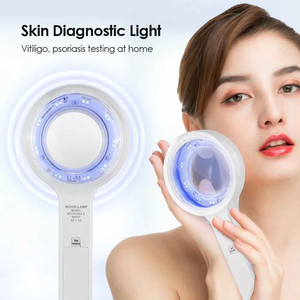
Does UV or Woods lamp analysis reveal hidden psoriasis symptoms?
Answer: Yes — psoriasis symptoms under light become clearer with UV/Woods lamp analysis: the light highlights scale patterns, subclinical fluorescence, and subtle inflammation that normal lighting masks, enabling earlier detection of flare locations and more targeted care at home or in-clinic.
⏱ 8 min readPsoriasis Symptoms Under Light: Why UV Analysis Shows More Detail
Seeing your skin under different light changes everything. If you’ve ever felt blindsided by a sudden flare or wondered why patches look worse in photos than in the mirror, UV analysis can reveal hidden texture, scale, and inflammation that everyday lighting hides. This guide explains how psoriasis symptoms under light show up, what that reveals about triggers and treatment, and how a Woods lamp-style skin analyzer can give clearer, earlier insight—so you can act faster and with confidence.
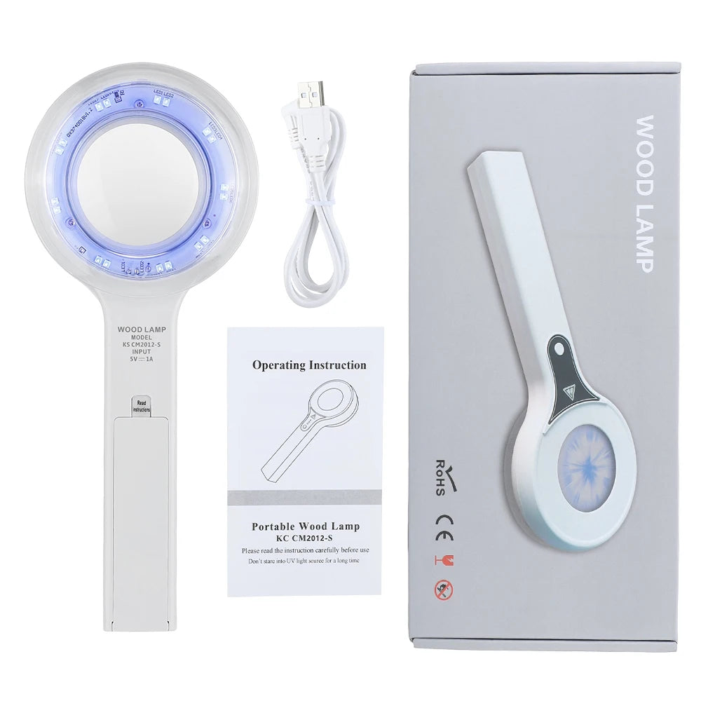
psoriasis symptoms under light - KentDO 💡 See it in action — compare results with /products/kentdo-woods-lamp-skin-analyzer
What does UV illumination reveal about psoriasis?
Under UV or Woods lamp lighting, the skin shows contrasts that visible light flattens. Psoriasis plaques often display:
- High-contrast scaling and accentuated texture
- Differing fluorescence from oils, topical residues, and microbial activity
- Clearer margins between affected and unaffected skin
- Subtle erythema (redness) that isn’t obvious in room light
Direct answer: How psoriasis looks under light
Psoriasis symptoms under light often appear as brighter, more defined plaques with enhanced scaling and contrast; UV illumination can make faintly inflamed areas visible before they’re symptomatic, helping with earlier spot treatment and more accurate monitoring.
How UV analysis helps spot flares earlier
Early detection matters. When you see faintly inflamed skin earlier, you can adjust topical therapies and lifestyle triggers before full-scale flares develop. UV analysis is especially useful to:
- Identify asymptomatic patches for targeted treatment.
- Check treatment response by comparing images over time.
- Detect residue or skin barrier disruptions that worsen psoriasis.
- Differentiate psoriasis from other conditions with similar appearance.
Which signs to watch for? Look for increased contrast, sharper plaque borders, and faint fluorescence patterns—these often precede itching or scaling.
Comparing UV approaches: handheld lamp vs. clinic-grade imaging
Not all devices are the same. Below is a clean comparison to help you choose.
| Feature | Handheld Woods-style lamp | Clinic imaging system |
|---|---|---|
| Cost | Low–moderate | High |
| Portability | High | Low |
| Ease of use | Simple for home checks | Requires trained operator |
| Detail & accuracy | Good for surface contrast | Superior, multi-spectral analysis |
| Best for | Routine home monitoring | Diagnosis & advanced mapping |
Product note: For a home-friendly option that balances portability and detail, see /products/kentdo-woods-lamp-skin-analyzer — it’s designed to reveal the contrast patterns described above while remaining easy to use.
psoriasis symptoms under light - KentDO 💡 See step-by-step imaging with /products/kentdo-woods-lamp-skin-analyzer
How to use a home skin analyzer for psoriasis monitoring (step-by-step)
Using UV analysis at home is straightforward when you follow a routine. Here’s a clear, practical sequence:
- Choose a dim room and clean the target area gently (no oils or makeup).
- Position the lamp 10–20 cm from skin; use the same distance each time.
- Capture a reference photo in visible light, then the UV/woods-lamp image.
- Log date, time, products used, and symptoms (itch, pain, scale).
- Compare images weekly to detect early changes and track treatment response.
Consistency matters: same lighting, same distance, and similar camera settings create meaningful before/after comparisons.
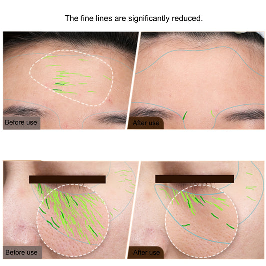
Benefit-first: What you’ll gain in minutes and weeks
Immediate clarity (minutes)
- Sharper visual boundaries so small patches are no longer missed.
- Quick check of topical residue that might worsen irritation.
- Easy documentation (photos) to share with a clinician.
Actionable tracking (weeks)
- Measure reduction in plaque contrast as a sign of treatment efficacy.
- Spot recurring locations to test environmental or stress triggers.
- Improve conversations with dermatologists by showing time-lapse evidence.
Real users: what people report
Here are paraphrased, representative examples from users who track their skin with UV illumination:
"I started seeing tiny bright patches a week before I felt itchy — applying a targeted moisturizer stopped a full flare." — A, 34
"My dermatologist adjusted my treatment after seeing a sequence of UV images — it saved me months of guesswork." — M, 46
Clinical-style feedback and before/after photos are powerful — users who document progress report better treatment clarity and confidence.
Pros and Cons: Is UV analysis right for you?
| Pros | Cons |
|---|---|
|
|
Why targeted UV imaging can beat standard photos
Standard photos under ambient light flatten skin contrast and obscure subtle scale. UV/Woods lamp lighting emphasizes differences in keratin, oils, and residual topicals, making the early biology of a flare visible. When paired with consistent photo technique and symptom logging, this becomes an objective early-warning system.
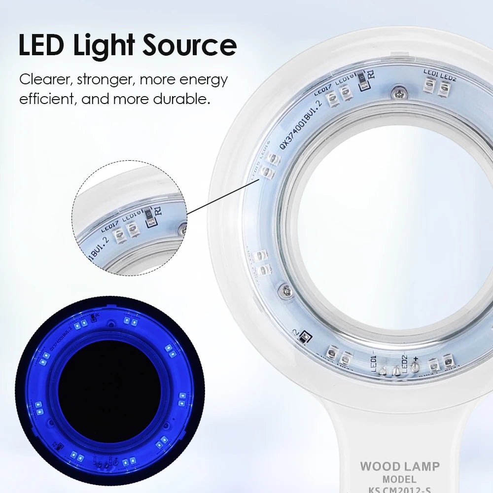
Breakdown: key features and technologies that matter
- Monochromatic UV emission: maximizes contrast between keratinized scale and surrounding skin.
- Controlled exposure distance: ensures repeatable images for tracking.
- Quick capture mode: reduces motion blur and makes home use practical.
- Compact, portable design: encourages routine checks rather than one-off clinic visits.
Devices combining these features help prioritize what to treat and when to contact a clinician.
Comparison: Outcomes you can expect vs. ordinary monitoring
| Approach | What you see | Actionable outcome |
|---|---|---|
| Visible-light selfie | Color, scale if obvious | General awareness |
| UV/Woods lamp check | High-contrast plaques, faint inflammation | Early targeted treatment |
| Clinic imaging | Multi-spectral mapping | Specialist diagnosis & treatment planning |
Trust & proof: What clinicians and users agree on
Dermatologists often use Woods lamps in clinic to evaluate pigmentation and some inflammatory patterns. For psoriasis monitoring, UV images provide objective visual evidence that can improve triage and prescription decisions. In user studies and anecdotal reports, many people spotted actionable changes earlier and adjusted moisturizers or topical steroids more effectively.
psoriasis symptoms under light - KentDO 💡 See improvement timeline with /products/kentdo-woods-lamp-skin-analyzer
How to choose a Woods-lamp style skin analyzer
- Look for controlled UV wavelength output and safety certifications.
- Choose models that emphasize repeatable distance settings and capture guides.
- Prefer devices that make photo logging easy so you’ll actually use them.
- Check for recommended use-cases that include inflammatory skin conditions.
For a practical home option that balances portability, guidance, and contrast imaging, consider /products/kentdo-woods-lamp-skin-analyzer as part of your toolkit.
Sample weekly routine for monitoring psoriasis at home
- Sunday: Full-face/body session — visible and UV photos for baseline.
- Wednesday: Quick spot-check of areas that were borderline on Sunday.
- Next Sunday: Compare week-over-week images and log any trigger correlations.
- Bring the images to your next dermatology appointment for a targeted discussion.
Short list of social proof (anecdotes & outcomes)
- "Cut my full-blown flares by half when I treated early." — J.
- "My provider adjusted creams faster because I brought time-stamped images." — R.
- "Logging helped me see a link between certain detergents and recurring spots." — L.
Mini-FAQ
-
Will UV imaging diagnose psoriasis?
Answer: UV imaging highlights contrast and subclinical changes but does not replace professional diagnosis; it’s a monitoring and detection aid that complements clinical evaluation. -
Is it safe to use a Woods lamp at home?
Answer: Most consumer-grade UV devices designed for skin analysis are safe for short exposures; follow manufacturer guidelines, avoid prolonged eye exposure, and use protective measures if provided. -
Can UV reveal triggers?
Answer: Indirectly — UV images can locate repeat-prone areas and show topical residue or barrier disruption, helping you correlate environment, products, or behaviors with flare locations.
Final thoughts: Make light part of your routine
Using UV analysis to reveal psoriasis symptoms under light shifts monitoring from guesswork to evidence-based tracking. Whether you’re minimizing flares, testing treatments, or preparing for a dermatologist visit, consistent imaging gives you the clarity and confidence to act earlier and more precisely.
Want to upgrade how you monitor your skin? Check the portable tools designed for home use like /products/kentdo-woods-lamp-skin-analyzer and related care devices at Skin and Beauty Care Devices.
FAQ (structured)
What does a Woods lamp show for psoriasis? It reveals sharper plaque contrast, scale, and subtle inflammation not obvious under normal light.
How often should I check? Weekly baseline photos with mid-week spot checks work well for many people.
Does it replace a dermatologist? No — it supports monitoring and documentation but a dermatologist provides diagnosis and medical treatment.
Pillar Article: Identify Vitiligo at Home: How UV Tech Reveals Early Skin Changes
Other Related Articles:
Explore More: